 English (UK)
  Русский (РФ)
 1. X-ray photoelectron spectroscopy (XPS) and X-Ray photoelectron imaging (XPi)
The method of photoelectron spectroscopy is a modern method of investigation of occupied electronic states in solids. It is based on the phenomenon of the photoelectric effect: electron in a solid is optically excited by a photon to the unoccupied state. Photoelectron spectroscopy of core levels enables to obtain quantitative information about elemental and chemical composition of the surface area of the samples. Method of elemental mapping of surface is used for study elemental and chemical composition of samples with lateral resolution, the implementation of this method is possible due to the presence of special microchannel plate, which allows to analyze the emitted photoelectrons from a solid body with a spatial resolution.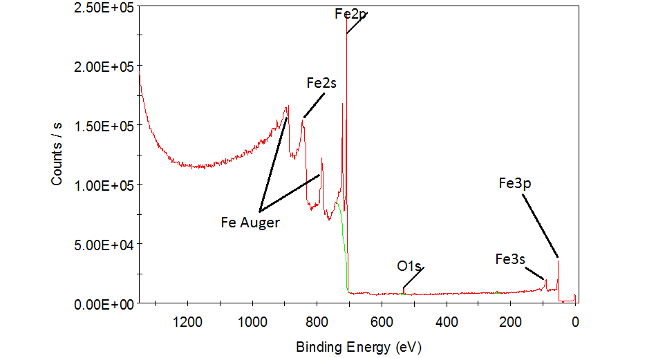
The example of survey XPS-spectrum (steel 09G2S)
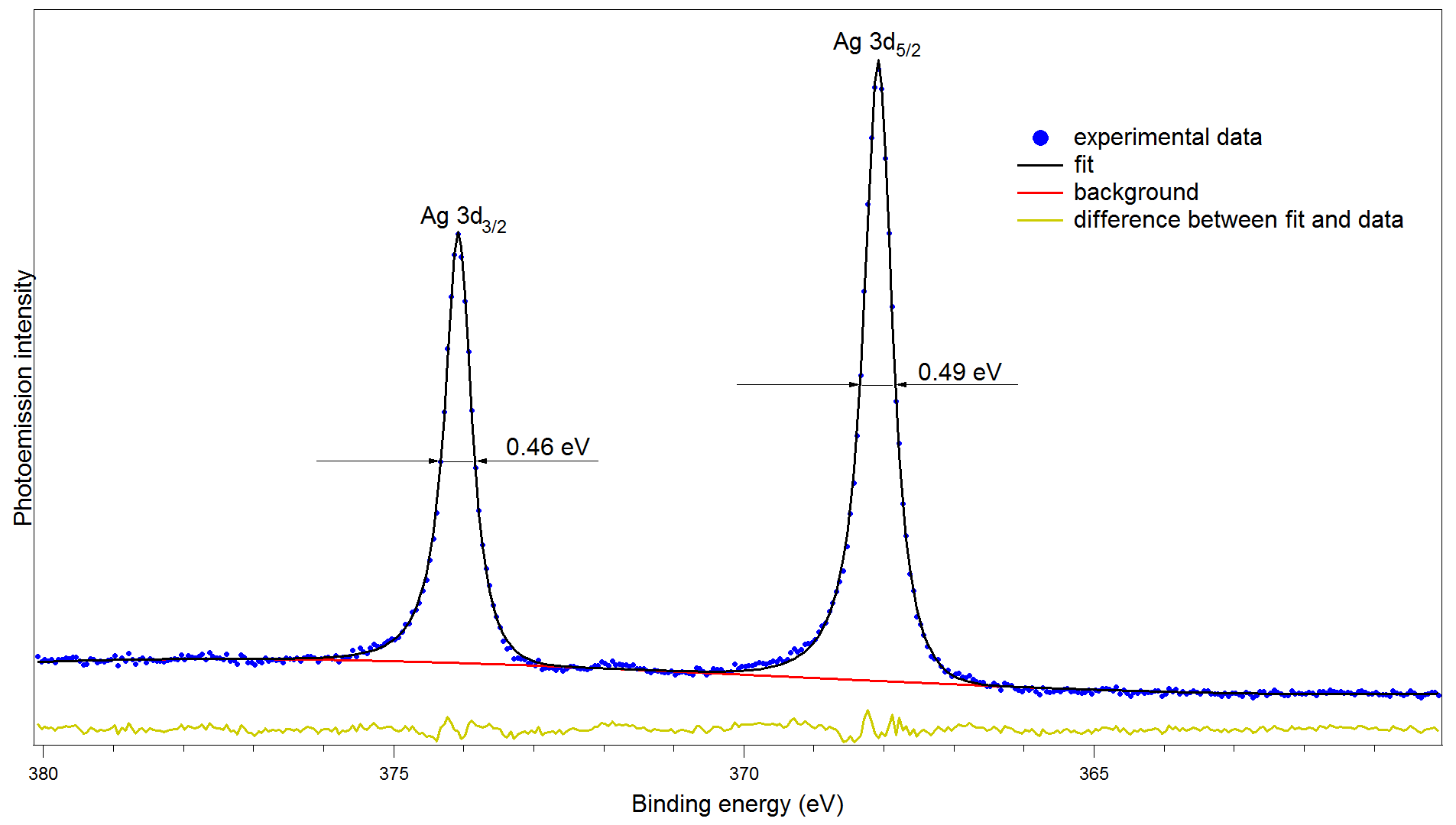
The doublet of the 3d-level of silver, approximated by the convolution
of the Gaussian and Lorentz functions, the full width at half maximum is 0.46 and 0.49 eV.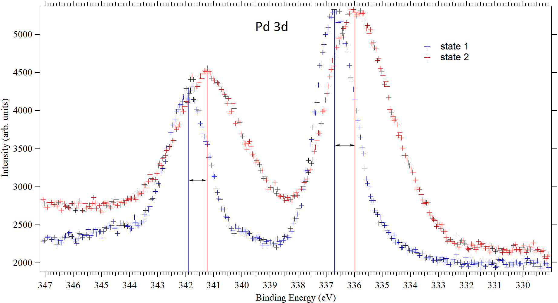
Core levels of Palladium in different states, shift=0.7 eV
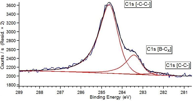
The decomposition of core level C1s into components (using as example B–graphene/Ni(111)/W(110) system)
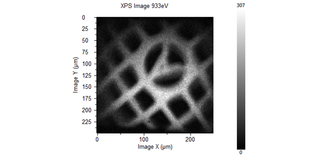
XPS-imaging of copper on the test sample surface
Escalab 250Xi Nanolab (without imaging) Univer-M (without imaging) 2. Spin and angle resolved photoelectron spectroscopy (SARPES) of valence band
The method of photoelectron spectroscopy with angular resolution is widely used for measuring of dispersions and symmetry of electron energy bands of solid state.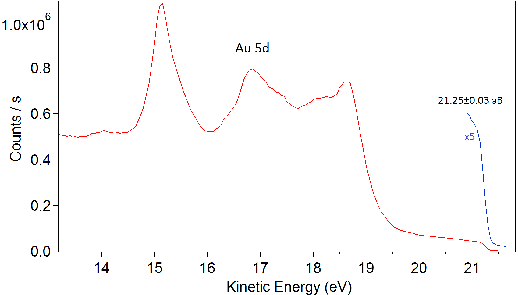
Photoelectronic valence band spectrum of polycrystalline gold HeI (21.2 eV)
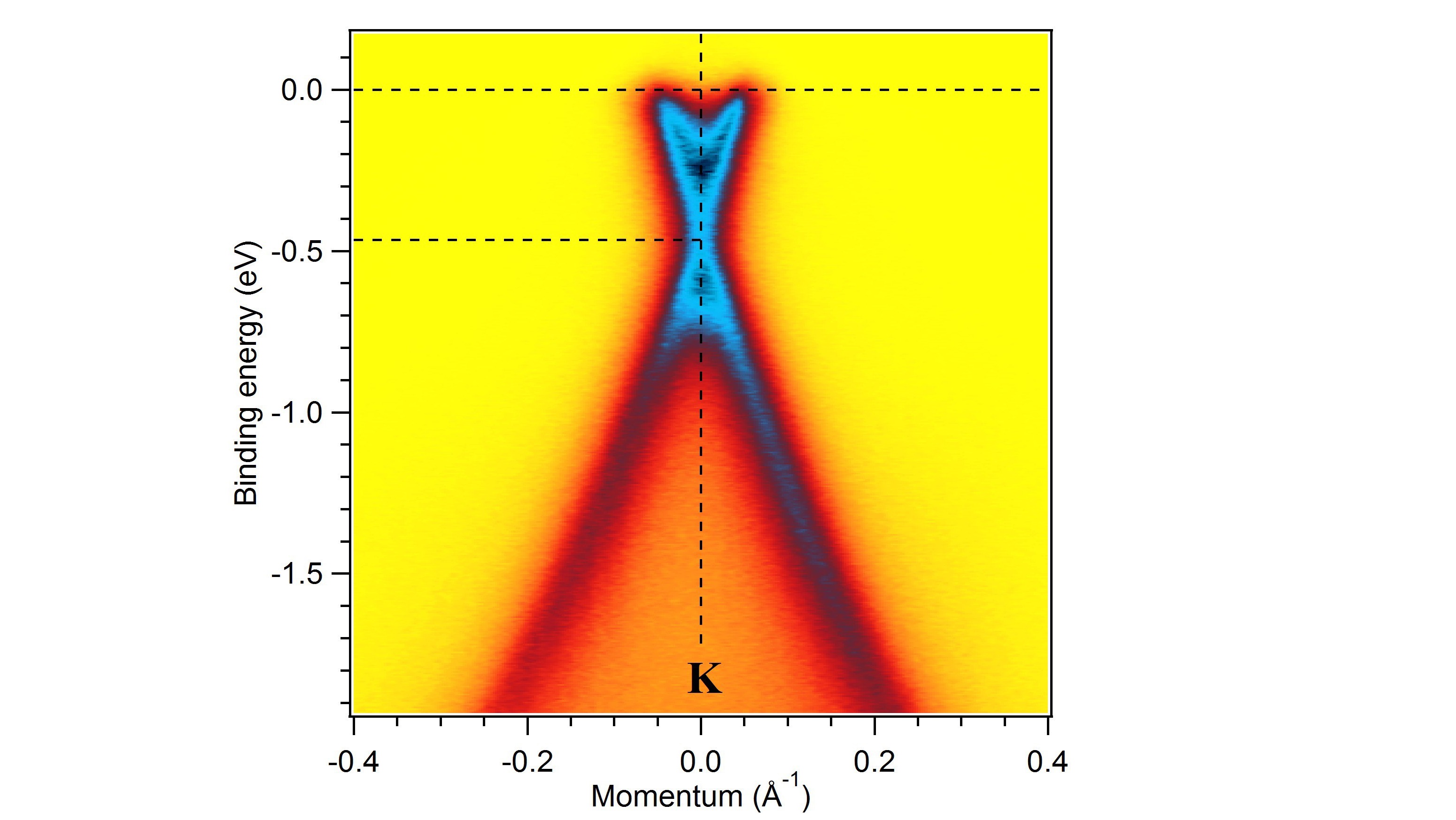
ARPES-image of band structure of graphene on SiC (Dirac cone)
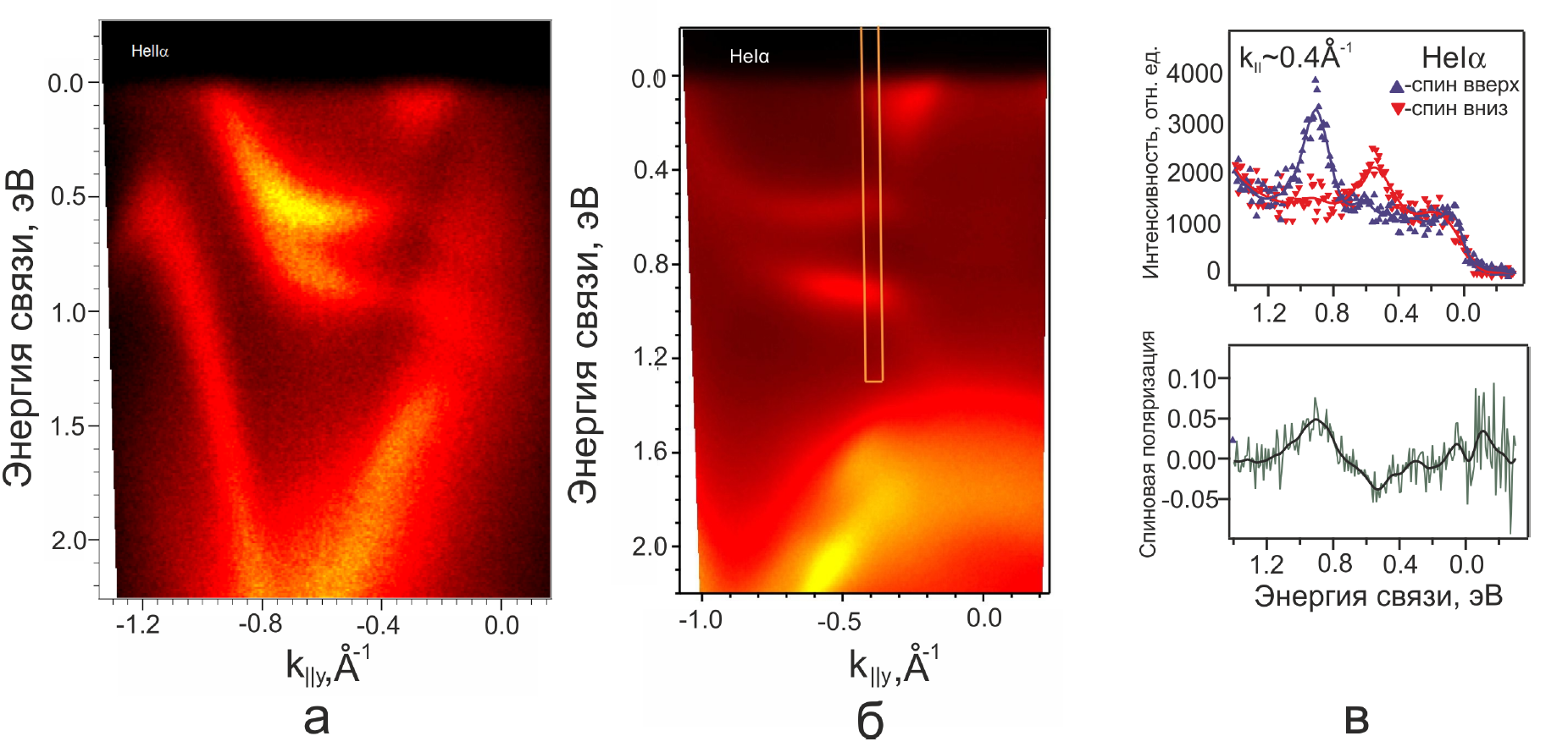
Photoelectronic spectra with spin resolution of system 1 ML Au / W(110) ГS-direction of Brillouin zone: а) photon energy = 40.8 eV, б) photon energy = 21.2 эВ, в) spin-resolved spectrum and its spin polarization, photon energy = 21.2 eV
Escalab 250Xi (Ultraviolet PES without angle and spin resolution) Nanolab Univer-M 3. Low energy electron difraction (LEED)
Method of LEED provides information about single crystal structure of the sample surface.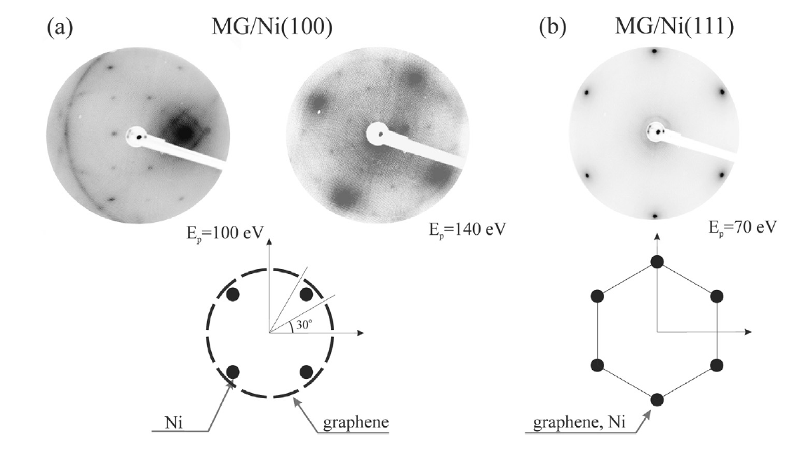
Graphene LEED on: (a) Ni(100), Ep = 100 eV, (b) Ni(111), Ep=110 eV.
Escalab 250Xi Nanolab Univer-M 4. Auger electron spectroscopy (AES) and scanning auger electron mapping (SAM)
The origin of Auger electron spectroscopy (AES) is measurement of energy and intensity of Auger electrons emitted from the sample surface when it is bombarded with a beam of electrons. An important feature of the Auger electron spectroscopy is its sensitivity to chemical state of analyzed elements on the surface. The chemical state of elements of the sample affects the shape and position of features of the spectrum of the Auger electrons.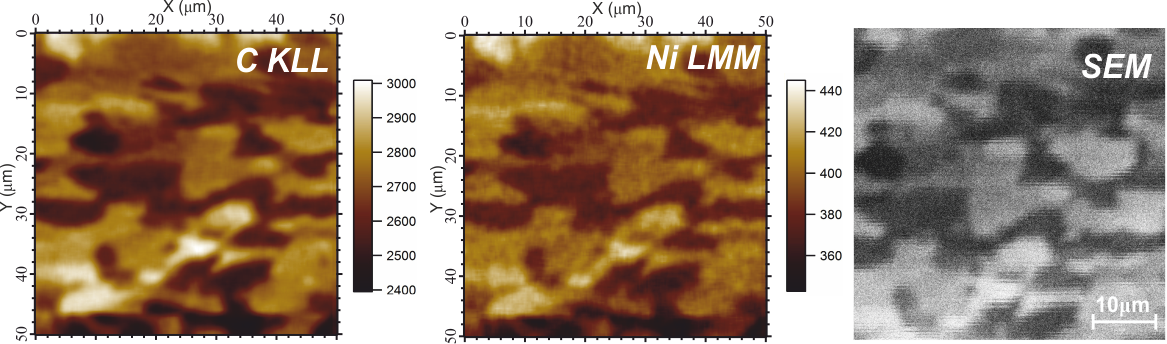
Auger maps (C KLL and Ni LMM lines) and SEM image (50×50 μm) of the sample after heating of system Ni/ВОПГ up to 310°C
Escalab 250Xi Univer-M 5. Scanning electron microscopy (SEM)
The method allows to obtain an image of the sample surface by scanning with focused electron beam (up to 95 nm and 10 keV) with simultaneous recording of low-energy secondary electrons, excited by this beam.Escalab 250Xi Univer-M 6. Ion scattering spectroscopy (ISS)
Ion scattering spectroscopy (ISS) is a technique in which a beam of primary ions is scattered by a surface. The kinetic energy of scattered ions is measured. Energy losses depend on the relative masses of the surface atoms and ions, thereby measured spectrum contains information about elemental composition.Escalab 250Xi 7. Reflected electron energy loss spectroscopy (REELS)
Electron energy-loss spectroscopy is a kind of electronic spectroscopy, when investigated surface irradiated by electrons with a narrow range of energies, and losses of energy of inelastically scattered electrons are recorded. Distribution of electron energies carries information about energy loss due to excitation of vibrational states, plasmons, deep levels and interband transitions.Escalab 250Xi 8. Scanning tunneling microscopy (STM) and scanning tunneling spectroscopy (STS)
These methods allow to obtain images of surface with atomic resolution, energy spectra of occupied and unoccupied states, distribution of work function and local density of states with high lateral resolution.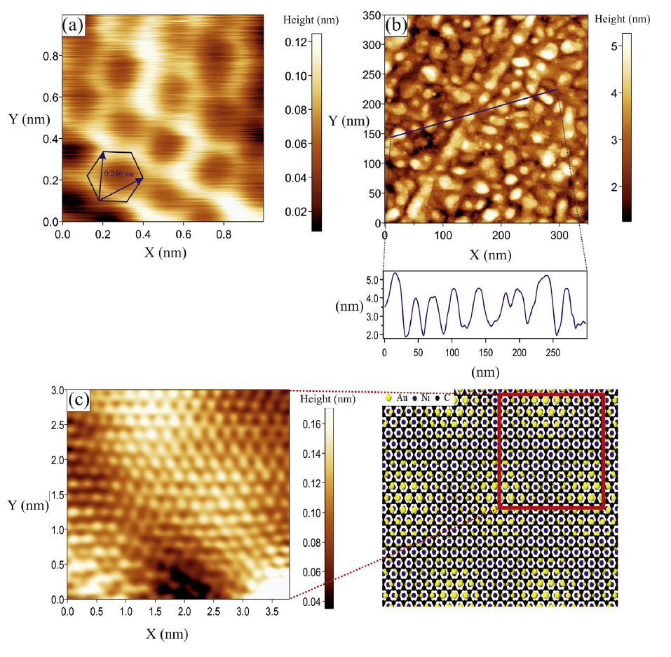
STM image of graphene-containing system surface, recieved by constant tunnel current: (а) graphene, synthesized on Ni-substrate; (b) gold, dusted on graphene-Ni system; (c) system after heating up to 310°C. Right peace shows model, wich demonstrates Moire pattern, arising because of overlapping of a gold monolayer and a graphene lattice.
Nanolab 9. Atomic force microscopy (AFM) (contact and non-contact modes)
Nanolab 10. Reflectron time-of-flight mass spectrometry (TOF-MS)
Time-of-flight mass spectrometry (TOFMS) is a method of mass spectrometry in which an ion's mass-to-charge ratio is determined via a time measurement. Using a reflector leads to a significant increase in resolution time-of-flight devices in compare with linear increases spectrometers and to an increase of mass determination accuracy. Selection of ionization source depends on substance state before ionization. Ionization is possible by electron impact or by laser radiation (photoionization).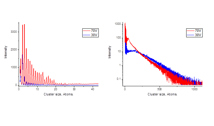
Mass spectra obtained in a reflectron in the supersonic beam ionization of xenon clusters by an electron beam with an energy of 30 and 70 V
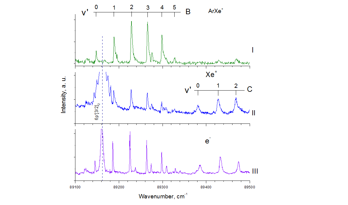
I - (2+1) photoionization spectrum of ArXe molecules in the region of the atomic state 6р'[3/2]2 upon registration of (-) ArXe + ions;
II - (2+1) photoionization spectrum of ArXe molecules in the region of the atomic state 6р'[3/2]2
upon registration of Xe+ ions;
III - CIS spectrum of ArXe molecules in the region Хе* 6р'[3/2]2, obtained at a fixed energy on a photoelectron spectrometer of 4.515 eV and when scanning laser radiation.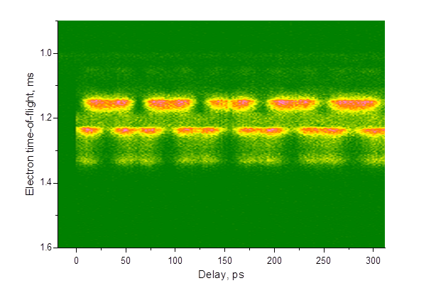
Larmor precession of excited xenon atoms in the magnetic field of the bottle, observed in the Pump-Probe experiment. Pumping by two-photon resonant excites xenon atoms into the state 5p5(2P°3/2)6p2[3/2]2. The probe pulse 795nm, 50fs ionizes these states two-photonly through intermediate levels.
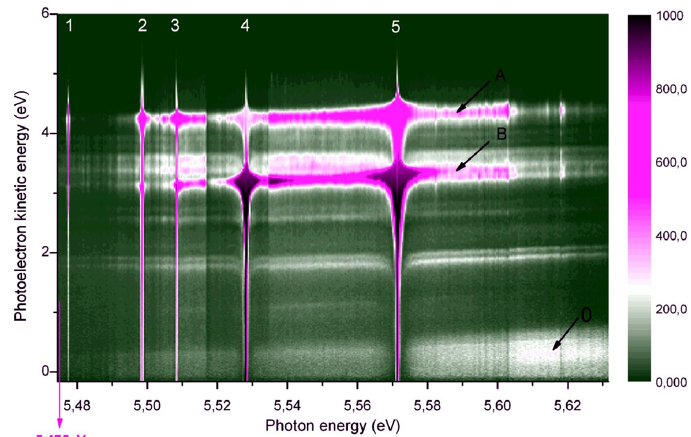
Photoelectron spectra of a supersonic molecular beam containing xenon clusters. The electron energy spectrum is recorded by a time-of-flight electron spectrometer of the "magnetic bottle" type. 0 - direct two-photon ionization of xenon clusters; A and B are the bands formed by three-photon ionization of xenon atoms, corresponding to two states of xenon ions Хе + 2Р1 / 2 и Хе + 2Р3 / 2.
Univer-М 11. Thermal desorption spectroscopy (TDS)
Essense of the method is measuring composition of desorbed gas from sample heated in a vacuum in dependence on the temperature. The method allows to determine binding energy of adsorbed atoms and molecules (or activation energy of desorption), amount of surface coverage by adsorbed substances, order of kinetics of adsorption process.Univer-М